Light Element Ptychographical Imaging
 Electron Ptychographic Diffractive Imaging of Boron Atoms in LaB6 Crystals.
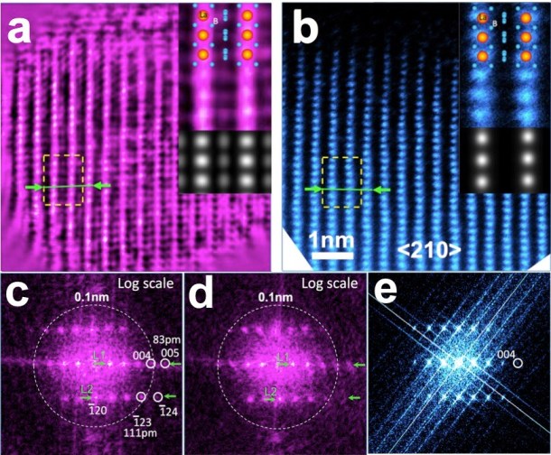
Here we show that electron ptychography can recover structural information for light elements located between heavy elements at atomic resolution and with high phase sensitivity, demonstrated in an experimental reconstruction of a LaB6 nanoparticle. This work was published in Scientific Reports.
3D Electron Ptychographical Imaging
Electron ptychographic microscopy for three-dimensional imaging
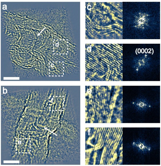
We reported a combination of electron ptychography with the inverse multislice method. Depth sectioning of a nanostructured material into slices with 0.34 nm lateral resolution and with a corresponding depth resolution of about 24-30 nm was demonstrated. This 3D imaging method has potential applications for the 3D structure determination of a range of objects, ranging from inorganic nanostructures to biological macromolecules. This work was published in Nature Communications.
Ptychography with Hollow Pixelated Detector (5D STEM)
Hollow Electron Ptychographic Diffractive Imaging
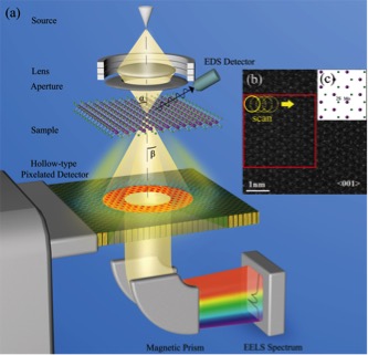
We report a method for quantitative phase recovery and simultaneous electron energy loss spectroscopy (EELS) analysis using ptychographic reconstruction of a data set of “hollow” diffraction patterns. This has the potential for recovering both structural and chemical information at atomic resolution with a new generation of detectors. This work was published in Physical Review Letters.
Low-dose Ptychographic Imaging
Low Dose Ptychography
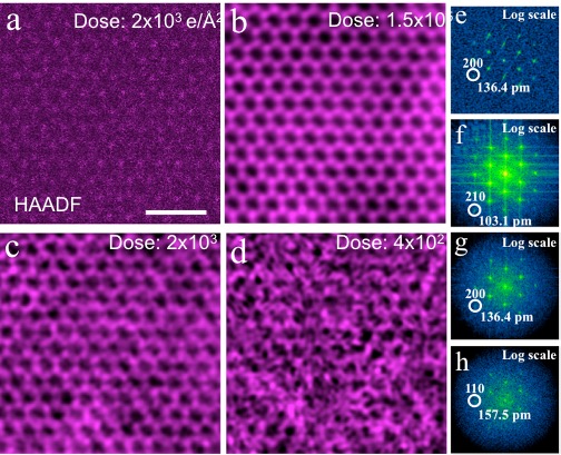
To date studies have been mainly limited to radiation-resistant samples as the electron dose required to record a ptychographic dataset is too high for use with beam-sensitive materials. Here we report defocused electron ptychography using a fast, direct-counting detector to reconstruct the transmission function, which is in turn related to the electrostatic potential of a two- dimensional material at atomic resolution under various low dose conditions. Finally, the parameters reported here should provide guidelines for ptychography of beam-sensitive samples including bio- logical materials. This work was published in Scientific Reports.
Low-dose High Contrast Cryo-Ptychography for Bio-Imaging
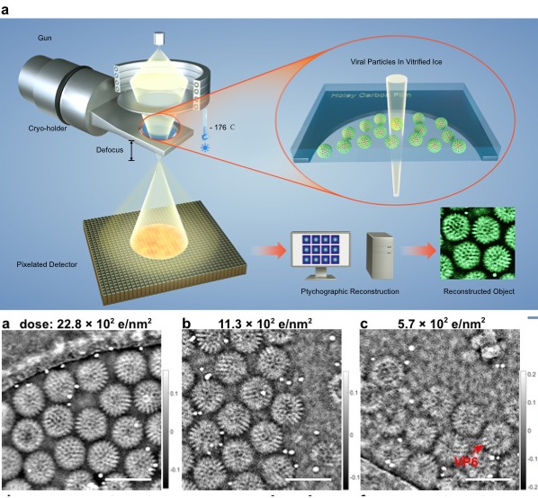
Cryo-Ptychography
Cryo-electron microscopy is an essential tool for high-resolution structural studies of bio- logical systems. This method relies on the use of phase contrast imaging at high defocus to improve information transfer at low spatial frequencies at the expense of higher spatial frequencies. Here we demonstrate that electron ptychography can recover the phase of the specimen with continuous information transfer across a wide range of the spatial frequency spectrum, with improved transfer at lower spatial frequencies, and as such is more efficient for phase recovery than conventional phase contrast imaging. We further show that the method can be used to study frozen-hydrated specimens of rotavirus double-layered particles and HIV-1 virus-like particles under low-dose conditions (5.7e/Å2) and heterogeneous objects in an Adenovirus-infected cell over large fields of view (1.14 × 1.14 μm), thus making it suitable for studies of many biologically important structures. This work was published in Nature Communications.
3D Ptychographic-Tomography
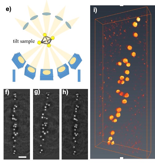
Ptychographic Tomography
Electron tomography has become a widely used and powerful technique for three dimensional (3D) structural analysis in both physical and biological sciences for decades. However, there are still remaining challenging in the areas, such as beam sensitive or low atomic number materials. In this work, we demonstrated that defocused-probe ptychography can produce 3D images from weak-scattering samples at low dose, based upon optical sectioning and tilt series approaches, respectively. This work was published in M&M 2019.
